Imatge:Nematocyst discharge.png
De Viquipèdia
No hi ha una versió amb una resolució més gran.
Nematocyst_discharge.png (480 × 371 píxels, mida de l'arxiu: 190 KB, tipus MIME: image/png)
| | Aquest arxiu és una càrrega compartida, extreta del projecte Wikimedia Commons i pot ser usada per altres projectes. Si voleu disposar de més informació sobre el fitxer, podeu visitar la pàgina original |
| Description |
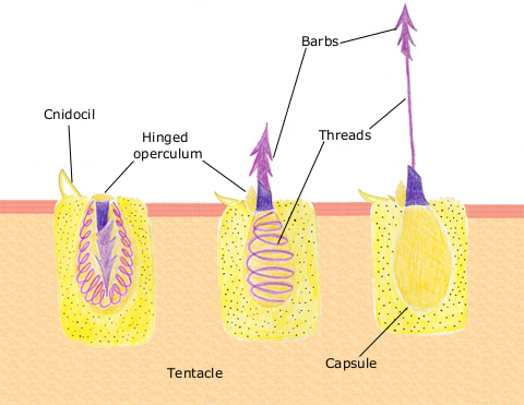
DescriptionThe diagram above shows the anatomy of a nematocyst cell and its “firing” sequence, from left to right. On the far left is a nematocyst inside its cellular capsule. The cell’s thread is coiled under pressure and wrapped around a stinging barb. When potential prey makes contact with the tentacles of a polyp, the nematocyst cell is stimulated. This causes a flap of tissue covering the nematocyst—the operculum—to fly open. The middle image shows the open operculum, the rapidly uncoiling thread and the emerging barb. On the far right is the fully extended cell. The barbs at the end of the nematocyst are designed to stick into the polyp’s victim and inject a poisonous liquid. When subdued, the polyp’s tentacles move the prey toward its mouth and the nematocysts recoil back into their capsules. |
|---|---|
| Source |
Originally from en.wikipedia; description page is/was here. |
| Date |
2007-04-11 (original upload date) |
| Author |
Original uploader was Spaully at en.wikipedia |
| Permission (Reusing this image) |
CC-SA; PD-USGOV-DOC-NOAA; PD-USGOV-NOAA. |
[edit] License information
| This file is licensed under Creative Commons ShareAlike 1.0 License |
 |
This image should be recreated using vector graphics as an SVG file. This has several advantages; see Commons:Media for cleanup for more information. If an SVG form of this image is already available, please upload it. After uploading an SVG, replace this template with template {{Vector version available|new image name.svg}} in this image. |
|
العربية | Български | Català | Česky | Dansk | Deutsch | English | Esperanto | Español | Français | 한국어 | Italiano | Magyar | Lietuvių | Nederlands | 日本語 | Polski | Português | Română | Русский | Suomi | Svenska | Türkçe | Українська | Tiếng Việt | 中文(繁體) | 中文(简体) | +/- |
|
| This image is in the public domain because it contains materials that originally came from the U.S. National Oceanic and Atmospheric Administration, taken or made during the course of an employee's official duties. |
|
[edit] Original upload log
(All user names refer to en.wikipedia)
- 2007-04-11 17:10 Spaully 480×371×8 (194868 bytes) Modified from: http://www.oceanservice.noaa.gov/education/kits/corals/media/supp_coral01b.html {{Information |Description=Nematocyst discharge process. |Source= Modified from [http://www.oceanservice.noaa.gov/education/kits/corals/media/supp_coral01b.html
Historial del fitxer
Cliqueu sobre la data/hora per veure el fitxer tal com era aleshores.
| Data/Hora | Dimensions | Usuari | Comentari | |
|---|---|---|---|---|
| actual | 19:29, 13 oct 2007 | 480×371 (190 KB) | Alison | ({{Information |Description===Description== The diagram above shows the anatomy of a nematocyst cell and its “firing” sequence, from left to right. On the far left is a nematocyst inside its cellular capsule. The cell’s thread is coiled under pressur) |
Enllaços a la imatge
Les següents pàgines enllacen a aquesta imatge:

