Imatge:Giardia lamblia SEM 8698 lores.jpg
De Viquipèdia
Mida d'aquesta previsualització: 479 × 599 píxels
Imatge en màxima resolució (700 × 876 píxels, mida de l'arxiu: 35 KB, tipus MIME: image/jpeg)
| | Aquest arxiu és una càrrega compartida, extreta del projecte Wikimedia Commons i pot ser usada per altres projectes. Si voleu disposar de més informació sobre el fitxer, podeu visitar la pàgina original |
| Description |
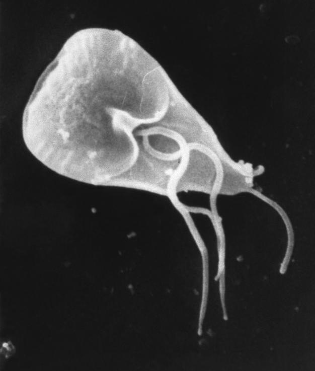
ID#: 8698 Description: This scanning electron micrograph (SEM) revealed some of the external ultrastructural details displayed by a flagellated Giardia lamblia protozoan parasite. G. lamblia is the organism responsible for causing the diarrheal disease "giardiasis". Once an animal or person has been infected with this protozoan, the parasite lives in the intestine, and is passed in the stool. Because the parasite is protected by an outer shell, it can survive outside the body, and in the environment for long periods of time. Cysts are resistant forms and are responsible for transmission of giardiasis. Both cysts and trophozoites can be found in the feces (diagnostic stages). The cysts are hardy and can survive several months in cold water. Infection occurs by the ingestion of cysts in contaminated water, food, or by the fecal-oral route (hands or fomites). In the small intestine, excystation releases trophozoites (each cyst produces two trophozoites). Trophozoites multiply by longitudinal binary fission, remaining in the lumen of the proximal small bowel where they can be free or attached to the mucosa by a ventral sucking disk. Encystation occurs as the parasites transit toward the colon. The cyst is the stage found most commonly in non-diarrheal feces. Because the cysts are infectious when passed in the stool or shortly afterward, person-to-person transmission is possible. While animals are infected with Giardia, their importance as a reservoir is unclear. Content Providers(s): CDC/ Janice Carr Copyright Restrictions: None - This image is in the public domain and thus free of any copyright restrictions. As a matter of courtesy we request that the content provider be credited and notified in any public or private usage of this image. |
|||
|---|---|---|---|---|
| Source | ||||
| Date | ||||
| Author |
Janice Carr |
|||
| Permission (Reusing this image) |
|
Historial del fitxer
Cliqueu sobre la data/hora per veure el fitxer tal com era aleshores.
| Data/Hora | Dimensions | Usuari | Comentari | |
|---|---|---|---|---|
| actual | 10:20, 31 maig 2006 | 700×876 (35 KB) | Patho | ({{Information| |Description=ID#: 8698 Description: This scanning electron micrograph (SEM) revealed some of the external ultrastructural details displayed by a flagellated Giardia lamblia protozoan parasite. G. lamblia is the organism responsible for caus) |
Enllaços a la imatge
Les següents pàgines enllacen a aquesta imatge:

